-
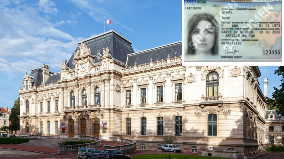
Where to find help with important admin in France
A website and phone helpline exist to help with all kinds of complex documents and processes
-
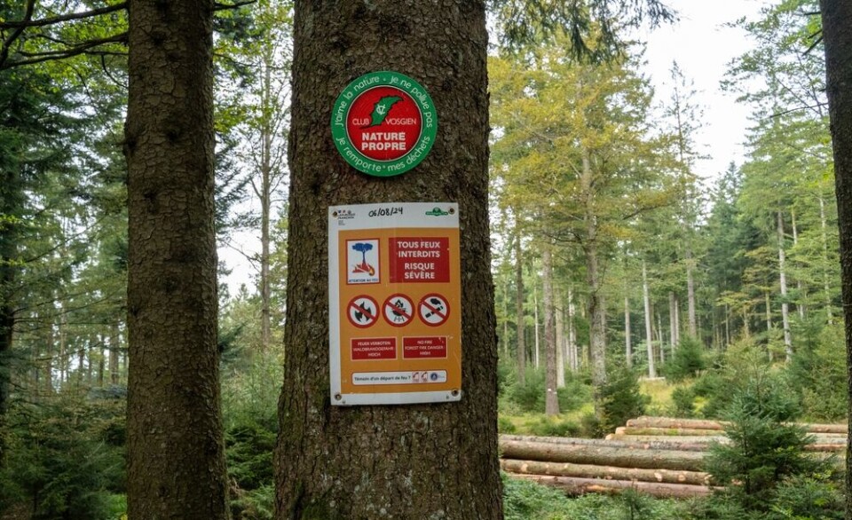
Bedbugs: French authority warns against banned insecticide
The product is still circulating despite being linked to four recent deaths
-
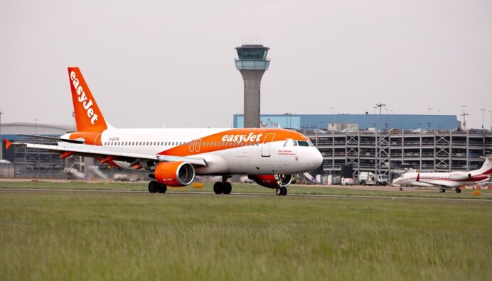
Air France increases flight prices again, by up to €50
For the second month in a row, rising kerosene prices increase flight ticket costs
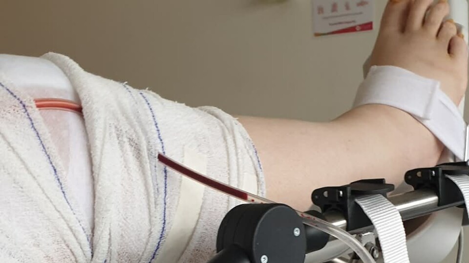
First child heart op using 3D scans
Surgeons perform pioneering keyhole surgery using ultrasound and X-ray pictures to avoid open-heart complications
THREE children have had pioneering keyhole heart operations in Toulouse with the surgeon being guided by a 3D computerised image on-screen.
The children, aged five, six and nine, were the first in Europe to have heart defects corrected using the Philips EchoNavigator software, which combines X-ray and ultrasound ‘transesophageal echocardiography’ 3D images to show the inside of the patient’s heart and body.
Using the equipment avoided having the children undergo difficult and demanding open-heart surgery – which can mean stopping the heart to allow the surgeon to operate - and possible complications with bleeding and infection.
The children were wakened just after the operation and were allowed home within 36 hours and without having any chest scars, which are disfiguring in later life.
Prof Philippe Acar, of Toulouse CHU, used the EchoNavigator and X-ray to guide him after inserting a catheter probe through a vein and, viewing images on screen, repaired a hole in five-year-old Nellie’s heart.
Working with Dr Sébastien Hascoet and Dr Khaled Hadeed then repeated the same delicate operation on a six-year-old boy and finally worked to close a hole in the upper chambers of the heart of a nine-year-old girl.
The technique has already been used on adults in other European hospitals and at the Henri-Mondor Hospital in Créteil (Val-de-Marne) but this is the first time it has been used on children.
About 40 other children are expected to undergo similar heart operations over the next two years as the technique is extended to other hospitals.
Traditional keyhole surgery uses a camera inside the chest cavity to show the surgeon where they are working but the 3D scan gave an exact view of the soft tissue Prof Acar was working on inside the heart.